INSIDE THE ROCK
Non-destructive by design
The Natural History Museum describes micro-CT as a non-invasive and non-destructive way to make 3D models of the inside and outside of specimens.
Time a scan doodlexrayz
00X-RAYS OF OBJECTS, NOT PEOPLE
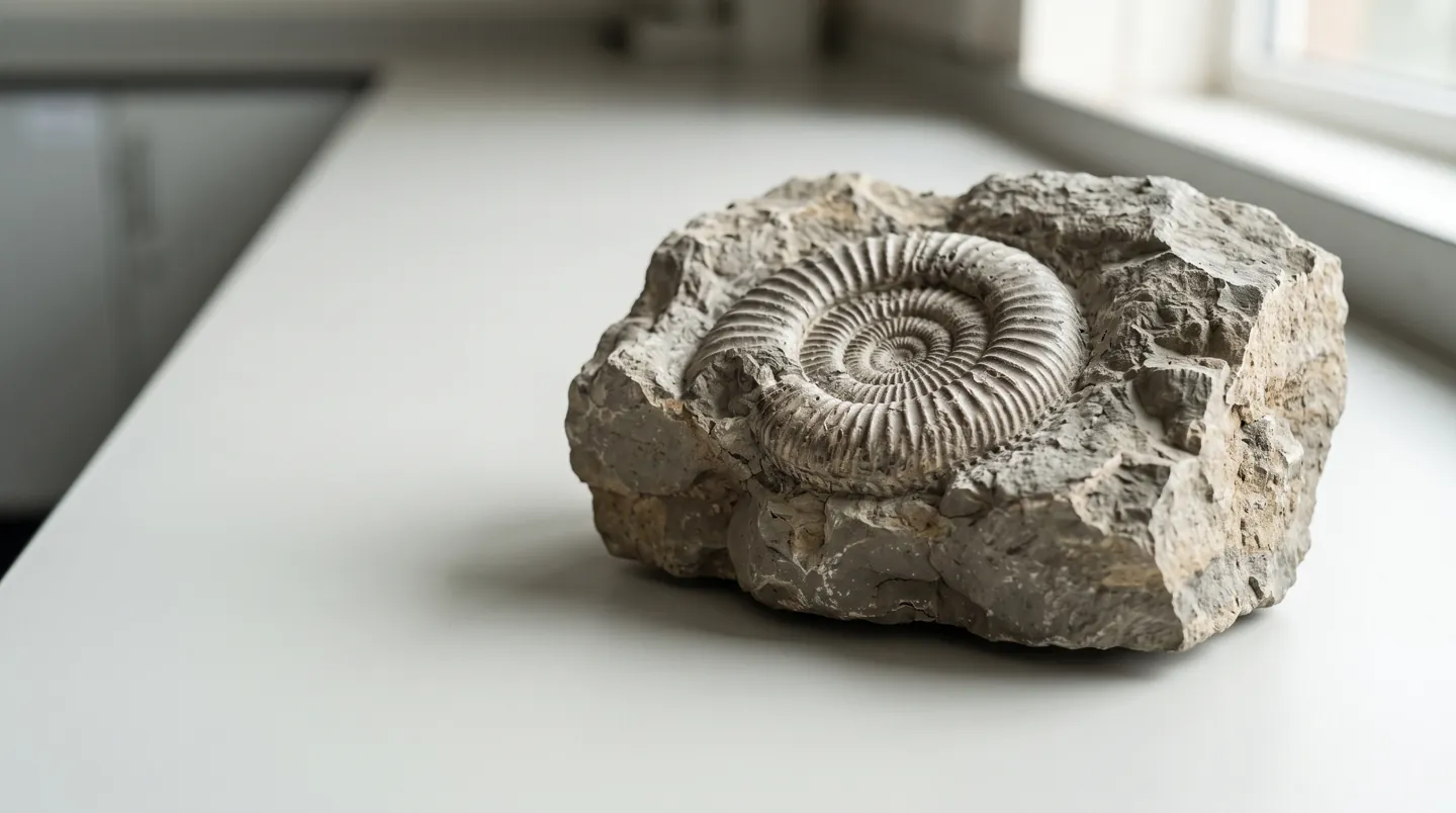
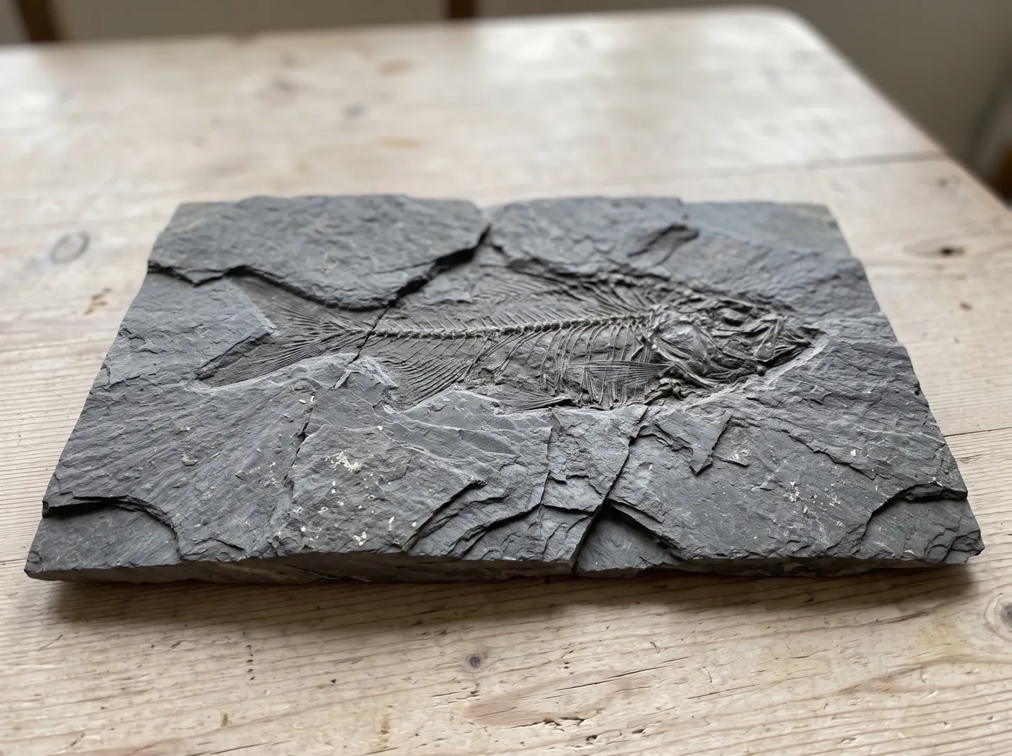
Some fossils never leave their stone.
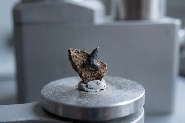
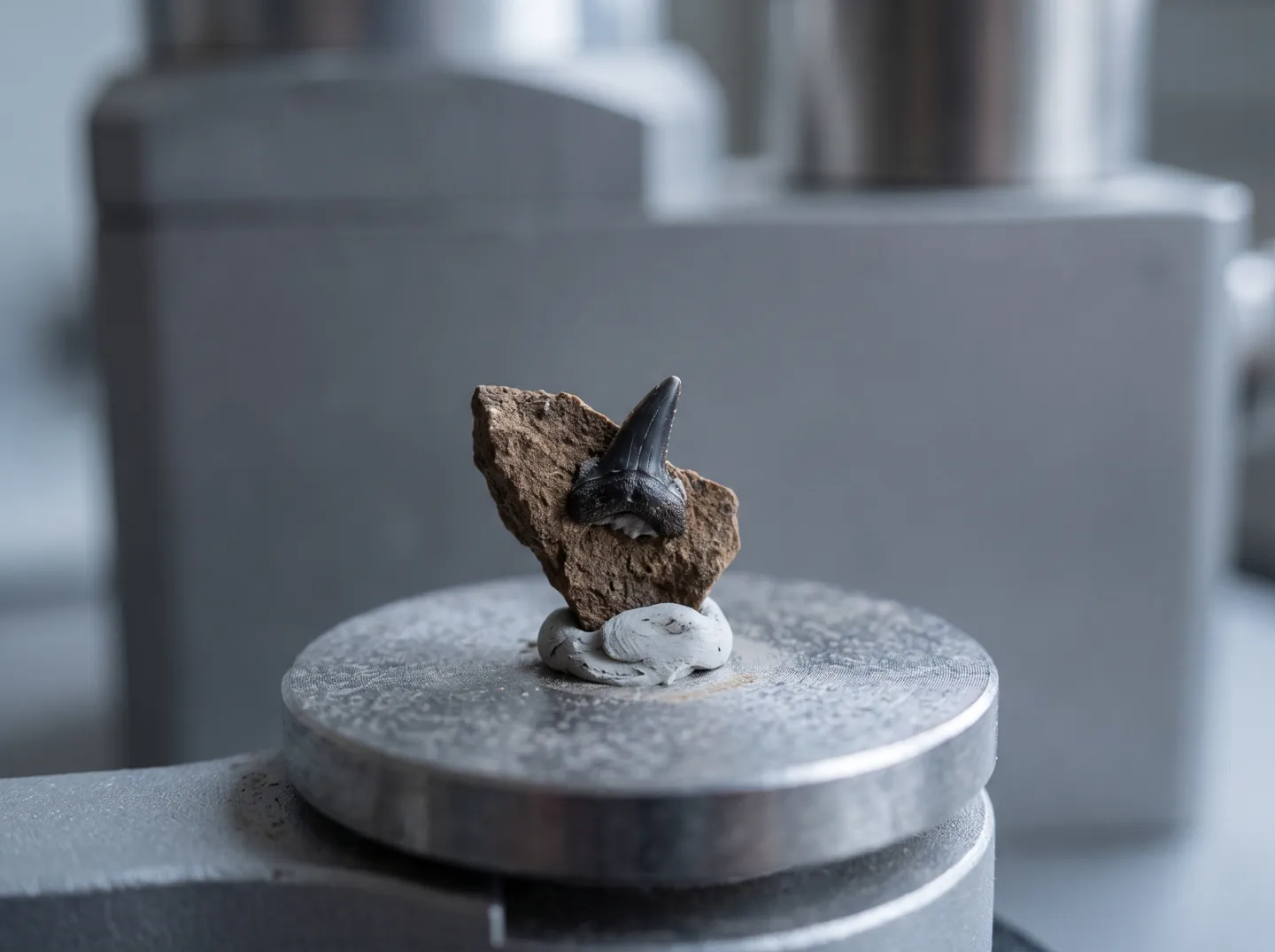
Micro-CT lets scientists see a fossil inside its rock without chipping a grain away. A museum worked example shows how a shark tooth about 315 million years old was scanned, sliced and freed on screen.
Time a scan01Principles
02THE SHORT ANSWER
CT measures how much X-ray gets through.
Software turns those views into slices you can explore.
INSIDE THE ROCK
The Natural History Museum describes micro-CT as a non-invasive and non-destructive way to make 3D models of the inside and outside of specimens.
Time a scanTHE SCAN
The fossil turns through 360° in a cone of X-rays while a detector records a radiograph every 0.05° to 0.1°, mapping how much X-ray passes through.
How slices are madeVIRTUAL PREPARATION
On screen, voxels are sorted by grey value into rock, tooth tissue and empty canals, so the fossil can be lifted out digitally.
The steps03A QUICK TIME CHECK
Pick a stage of making a virtual fossil. The bar shows how long it takes in the worked example, from the scan itself to preparing the fossil on screen.
Based on Abel, Laurini and Richter (2012), Palaeontologia Electronica, a micro-CT workflow using a Natural History Museum fossil shark tooth.
04TWO NUMBERS WORTH KNOWING
How small each 3D pixel was for the shark tooth,
and how big the detector that caught the X-rays.
0.009 mm
Size of each 3D pixel in the scan
The effective spatial resolution was 0.050 mm or coarser, because resolution is usually two to five times the voxel size. The best voxel size is about 1/2000th of the fossil's width or height.
VOXEL SIZE · SHARK TOOTH
2,000 × 2,000
Pixels across the detector panel
Each row of detector pixels becomes one slice. The tooth was scanned at 180 kV and 138 µA; a larger or denser fossil would need higher settings.
DETECTOR · MICRO-CT
Source: Abel, Laurini and Richter (2012). The fossil is Sphenacanthus hybodoides, a freshwater shark from Longton, Staffordshire, that lived about 315 million years ago.
05FROM SCAN TO VIRTUAL FOSSIL
The paper sets out a ten-step workflow. Grouped together, it comes down to four jobs after the scan.
Reduce noise, which makes scans grainy, and correct beam hardening, which makes the edges of a dense fossil look brighter than its centre.
Spread the fossil's grey values so that tooth tissue and rock are easier to tell apart.
Use a threshold, a region-growing tool and a mask to separate the fossil from the rock matrix around it.
Light and false-colour the model, animate it, then archive the slice stack so other researchers can use it.
06HOW SLICES ARE MADE
The word comes from the Greek tomos (slice) and graphein (to write). Software lines up the radiographs like the spokes of a bicycle wheel and back-projects them, smearing them across each other to build each slice from voxels, or 3D pixels.
Each voxel's grey value reflects how strongly the material absorbs X-rays, which depends mostly on density but also on thickness and atomic number. Allan Cormack and Godfrey Hounsfield shared the 1979 Nobel Prize for developing computer assisted tomography.
07Questions
It is X-ray computed tomography at a fine scale. The Natural History Museum describes it as a non-invasive, non-destructive technique that uses X-rays to create 3D models of the internal and external features of specimens.
CT is described as non-destructive: the fossil is X-rayed, not cut. Racicot (2017) adds that a scan becomes a lasting digital record that can be shared with researchers, museums and the public.
That is beam hardening. The X-ray beam mixes energies, and lower-energy X-rays are absorbed first, so the beam gets more penetrating as it passes through. A copper filter or later software correction can reduce it.
Mainly voxel size. Features smaller than a voxel can blur or disappear, and at very small voxel sizes noise becomes the limit. The fossil must fill the detector for the best magnification.
Abel and colleagues note that the first system, developed by Godfrey Hounsfield and Allan Cormack, went online in 1972 at Atkinson Morley's Hospital in London. The same principle is used for fossils today.
08Sources
ABOUT THIS NAME
doodlexrayz.com is part of the DoodleSale portfolio. It isn't in an auction right now, but you can ask about leasing it.